Unlocking Parkinson’s Mysteries: EEG and fMRI Synergy
In the quest to understand and combat Parkinson’s disease (PD), researchers are increasingly turning to advanced neuroimaging techniques. Among these, Electroencephalography (EEG) and Functional Magnetic Resonance Imaging (fMRI) stand out for their complementary strengths. This post explores how integrating EEG and fMRI enhances our understanding of PD. It offers insights into diagnosis, disease progression, and therapeutic interventions, demonstrating the value of combining these technologies.
Understanding EEG and fMRI
- Electroencephalography (EEG): EEG captures the brain’s electrical activity with high temporal resolution, detecting rapid neuronal events on the order of milliseconds. However, its spatial resolution is limited due to the diffusion of electrical signals through various brain tissues, making precise localization challenging.
- Functional Magnetic Resonance Imaging (fMRI): Conversely, fMRI measures brain activity indirectly by detecting changes in blood flow, known as the Blood Oxygen Level-Dependent (BOLD) signal. This method provides excellent spatial resolution, typically around 2–3 millimeters, allowing for precise mapping of active brain regions. However, fMRI’s temporal resolution is constrained by the slower hemodynamic response, which unfolds over several seconds, limiting its ability to capture rapid neuronal dynamics. This is particularly relevant in Parkinson’s research, where motor fluctuations and neural activity changes occur on a much shorter timescale. Understanding these rapid shifts is crucial for identifying biomarkers and improving therapeutic strategies.
The Power of Integration
Combining EEG and fMRI leverages their complementary strengths, offering a more comprehensive understanding of brain function. This integration allows researchers to achieve both high temporal and spatial resolution, facilitating the characterization of neural networks with greater precision. However, challenges such as data fusion and artifact management persist, necessitating advanced methodologies to effectively harness the full potential of simultaneous EEG-fMRI studies.
Advancements in Parkinson’s Research
- Functional Connectivity Alterations: Studies have demonstrated that PD patients exhibit reduced functional connectivity in primary EEG frequency bands, particularly alpha and beta rhythms. This reduction suggests early disruptions in neural communication pathways, potentially preceding observable spectral power changes. Such findings highlight the sensitivity of connectivity measures in detecting early-stage PD alterations.
- Impact of Pharmacological Intervention: Simultaneous EEG-fMRI studies have shed light on the effects of dopaminergic treatments, such as L-Dopa, on brain connectivity. Post-medication assessments reveal increased connectivity within motor networks, dorsal attention networks, and default mode networks. These changes underscore the modulatory role of dopamine on neural circuits and its therapeutic implications for PD symptomatology. Understanding these effects can guide personalized treatment approaches, optimizing dopaminergic therapies to enhance motor function and mitigate cognitive decline in patients with Parkinson’s disease.
- Advancements in Diagnostic Accuracy: The fusion of EEG and fMRI data, coupled with machine learning algorithms, has enhanced diagnostic precision in PD. For instance, employing the AdaBoost algorithm on combined EEG-fMRI datasets achieved a diagnostic accuracy of 93.45%, emphasizing the potential of multimodal approaches in early and accurate PD detection.
- Network Dynamics and Disease Progression: Investigations into the dynamic functional connectivity of PD patients have revealed abnormalities in brain network configurations. These disruptions correlate with motor and cognitive impairments, offering insights into the progression of the disease and potential biomarkers for monitoring therapeutic efficacy.
Spatial Resolution Changes in Parkinson’s Disease
Advancements in neuroimaging, particularly high-spatial-resolution diffusion MRI, have provided in vivo evidence of structural changes in the brains of PD patients. A study using diffusion tensor imaging (DTI) found significant microstructural alterations in the substantia nigra, correlating with disease severity. These findings suggest that diffusion MRI can serve as a biomarker for early detection and disease progression monitoring in PD. Notably, studies have observed lateral asymmetry in the substantia nigra, a region critical for motor control, suggesting that diffusion MRI is sensitive to such tissue changes in PD.
Dementia and Neuroimaging in Parkinson’s Disease
Cognitive decline, leading to Parkinson’s disease dementia (PDD), is a significant concern. Neuroimaging has revealed decreased cortical volume, increased white matter diffusion changes, and decreased resting metabolic activity in PD patients, which appear to begin prior to the onset of dementia.
Furthermore, heterogeneous neuroimaging findings in PDD have been linked to a common brain network centered on the hippocampus, a region essential for memory. This suggests that early neuroimaging assessments targeting hippocampal network integrity could aid in early diagnosis of Parkinson’s-related dementia. Additionally, understanding these network disruptions may inform the development of targeted interventions, such as neurostimulation or pharmacological treatments, aimed at preserving cognitive function in PD patients. This network-specific localization underscores the potential of neuroimaging in understanding and potentially predicting cognitive decline in PD patients.
Conclusion
The integration of EEG and fMRI modalities provides a robust framework for understanding the complex neural alterations in Parkinson’s disease. This multimodal approach facilitates early diagnosis, monitors disease progression, and evaluates treatment responses, thereby contributing to the development of targeted interventions and improved patient outcomes.
AI-generated medical infographics on Parkinson’s symptoms, treatment advances, and research findings; I hope you found this blog post informative and interesting. www.parkiesunite.com by Parkie
Image Prompt:
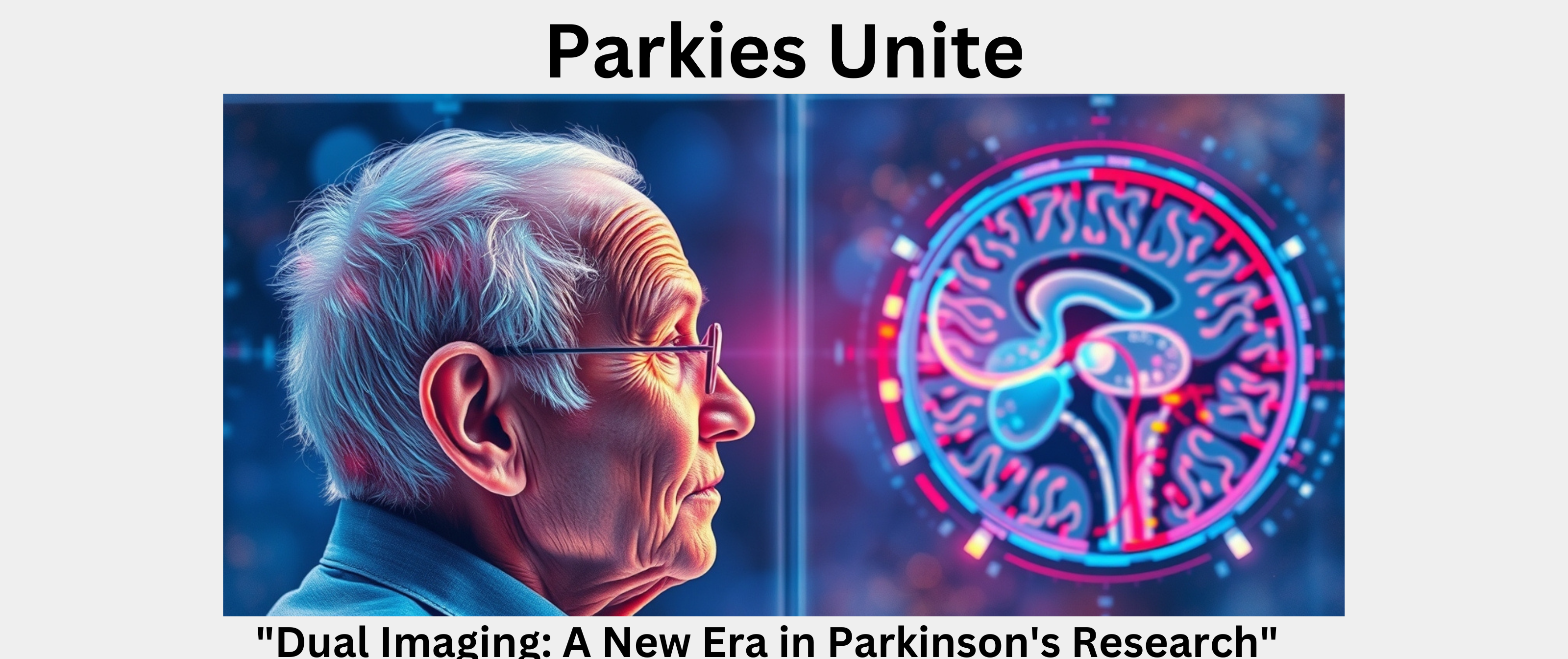
“A photo-realistic image depicting a person undergoing simultaneous EEG and fMRI scanning, highlighting the integration of electrical activity and blood flow measurements in the brain.”
Taglines:
- “Merging Brainwave and Blood Flow Imaging”
- “Dual Imaging: A New Era in Parkinson’s Research”
- “EEG-fMRI Synergy: Mapping Parkinson’s Pathways”
Negative Prompt:
“Malformed limbs, extra limbs, mutated hands, disfigured face, bad anatomy, malformed hands, text, lettering, captions, generating images with text overlays.”