Parkinson’s Brain Tremor Insights
Long-Form Blog Post :
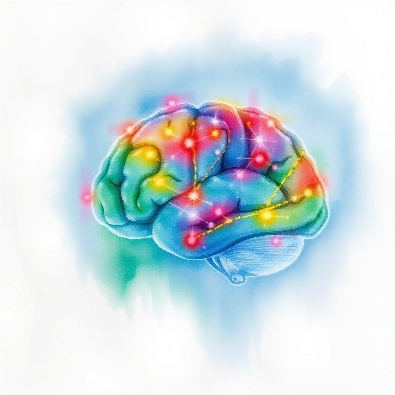
Understanding the evolving patterns of Parkinson’s disease symptoms requires looking closely at the ongoing neurodegenerative changes within the brain. Parkinson’s disease, driven by progressive dysfunction and death of dopaminergic neurons, impacts the delicate balance of motor control. As the disease advances, motor symptoms like tremors, rigidity, and bradykinesia often emerge and develop along distinct trajectories. To delve deeper into these complexities, researchers continue to explore structural changes in the brain’s gray matter regions and how they relate to symptom progression. This intersection of clinical observation and imaging-based investigation is critical for guiding future treatment strategies and improving patient outcomes.
One study featured in the journal Movement Disorders provides a prime example of such integrative research. Conducted by researchers in the Netherlands, this investigation drew on data from the Personalized Parkinson Project, a large prospective study of people diagnosed with Parkinson’s disease for no more than five years. In this project, 520 patients and 60 healthy controls underwent assessments designed to track changes in motor symptoms and correlate these shifts with MRI-based observations of brain structures over time.
At the outset (baseline), many patients exhibited bradykinesia, rigidity, and various forms of tremors: resting tremors, postural tremors, and kinetic tremors. Resting tremors typically occur when muscles are not in motion, while postural tremors appear when holding a position against gravity (such as outstretched arms), and kinetic tremors emerge during voluntary movements. Although commonly associated with Parkinson’s, tremors vary considerably: in some patients, tremors worsen over time; in others, they remain stable or even recede as the disease progresses.
After two years, it became evident that tremor progression patterns were quite different from those of bradykinesia and rigidity. The latter two symptoms tended to worsen consistently, reflecting the increasing difficulty in motor control that many patients face. In contrast, tremors — particularly kinetic and postural tremors — did not simply worsen. In fact, kinetic tremors were found to be less severe after two years, and postural tremors also showed a similar pattern. Resting tremors tended to stay relatively constant rather than progressing at the pace of other motor symptoms.
This unexpected trend set the stage for a more detailed examination of the underlying brain changes. Using MRI scans, researchers focused on regions implicated in Parkinson’s tremors: the thalamus, motor cortex, globus pallidus, and cerebellum. Over two years, progressive gray matter atrophy emerged in these areas, as well as throughout the whole brain. Interestingly, this atrophy was linked to the observed changes in tremors. Regions that underwent significant shrinkage were associated with reduced severity of kinetic and postural tremors. In other words, as the neural circuits supporting these tremors degenerated, they became less capable of producing the oscillatory firing patterns that give rise to action tremors.
However, while reduced tremor severity might seem like a positive development, the picture is more complex. The same overall gray matter atrophy correlated with progressive worsening of other motor symptoms, notably rigidity and bradykinesia. Areas of the brain that once modulated muscle tone and controlled movement speed became less effective as neuronal connections deteriorated. This illustrates a fundamental aspect of Parkinson’s disease: even as some symptoms like certain tremors may diminish, others often intensify, reflecting a shifting landscape of neurodegeneration and compensatory reorganization within the brain’s motor circuits.
These findings are further complicated by the interplay between disease-specific regions and overall brain health. Whole-brain analyses revealed that widespread cortical thinning correlated not only with reduced kinetic tremors but also with more severe bradykinesia and rigidity. Moreover, shrinkage in the hippocampus, a region critical for memory and learning, was associated with cognitive decline. Taken together, this suggests that the disease’s progression is not uniform. Instead, it affects distinct neural substrates differently over time, producing a complex picture of motor symptoms that do not all follow the same trajectory.
From a clinical standpoint, the implications are significant. For example, medication-resistant tremor is sometimes treated with surgical interventions such as deep brain stimulation (DBS) in earlier stages of the disease. If future research can reliably predict how an individual patient’s tremor severity will evolve, it could inform clinical decision-making. This would mean determining whether certain interventions are likely to remain effective or if a more aggressive approach may be needed earlier to manage tremor symptoms before they either fade or become especially troublesome.
Understanding why certain tremors dissipate as Parkinson’s disease evolves involves looking at the structure and function of the underlying neural circuits. Early on, abnormal synchronous firing between the basal ganglia, thalamus, and motor cortex can drive tremors. As these loops degrade due to neuronal loss and structural changes, they may lose their capacity to generate the rhythmic activity that manifests as tremor. In effect, the circuits required for sustaining action tremors break down. This loss is not an improvement in the disease state but rather the neural equivalent of machinery wearing out.
Meanwhile, bradykinesia and rigidity persist and worsen because the circuits that ensure fluid and efficient movement are heavily dependent on dopaminergic signaling and proper structural integrity. As dopamine production falters and anatomical atrophy intensifies, patients find it increasingly difficult to initiate movements or counteract muscle stiffness. The complexity arises because different symptoms emerge from distinct but interconnected neural substrates, each deteriorating at a unique pace.
From a research perspective, large-scale, longitudinal imaging studies like the Personalized Parkinson Project are invaluable. They help map how varying symptom trajectories relate to structural changes over time, providing essential clues to the underlying “circuit-level” mechanisms of Parkinson’s disease. Beyond simply acknowledging that neurodegeneration occurs, these efforts reveal how particular brain regions are tied to specific symptoms.
In the future, building robust predictive models based on MRI data, clinical assessments, and potentially other biomarkers may allow healthcare providers to anticipate symptom changes more accurately. This predictive capability could guide treatment strategies, ensuring that interventions are not just reactive but also proactive, addressing the needs of individual patients as their disease evolves.
In summary, this detailed interplay between atrophy in tremor-related brain regions and Parkinson’s symptoms such as tremor, rigidity, and bradykinesia underscores the complexity of neurodegeneration. While atrophy correlates with diminished action tremors, it simultaneously aligns with the progression of more disabling symptoms. Clinically, this complexity accentuates the importance of tailored interventions and the potential of using imaging studies to help guide patient-specific management. As more research refines our understanding, integrating structural data, clinical observations, and compensatory mechanisms may enable personalized approaches to treating Parkinson’s disease, ultimately improving quality of life for those affected.
“AI-generated medical content is not a substitute for professional medical advice or diagnosis; I hope you found this blog post informative and interesting. www.parkiesunite.com by Parkie”
Keywords : Parkinson’s disease, brain atrophy, tremor, MRI studies
DALL·E Prompt:
A delicate watercolor painting depicting interconnected brain regions fading at the edges, subtle neurons dissolving into soft hues of blues and greens, symbolizing Parkinson’s disease progression and evolving tremor patterns.